Abstract
Purpose. Type 2 diabetes occurs spontaneously in rhesus monkeys and shows an extraordinary similarity to human diabetes in clinical features and relative time course. The purpose of this study was to investigate clinically and histopathologically the ocular changes in these monkeys.
Methods. Ophthalmoscopic examinations were performed on aged normal and diabetic monkeys. Retinas from 16 diabetic monkeys and 6 nondiabetic monkeys were incubated postmortem for adenosine diphosphatase (ADPase) activity (labels viable retinal blood vessels) and flat-embedded in JB-4. Tissue sections were cut through areas of interest.
Rresults. Cotton-wool spots, intraretinal hemorrhages, and hard exudates in the macula were observed by ophthalmoscopy in some diabetic monkeys. Dot/blot hemorrhages, cotton-wool spots, and small nonperfused areas were the earliest histologically documented changes in the retinas. Large nonperfused areas extending from optic disc to midfovea were observed in four diabetic monkeys. Formation of small intraretinal microvascular abnormalities (IRMAs) and microaneurysms were associated with the areas of nonperfusion. There were apparent fluid-filled spaces in the outer plexiform layer in three of these maculas, suggesting macular edema. There was a significant correlation between the occurrence of retinopathy and hypertension (P = 0.037 for systolic pressure; P = 0.019 for diastolic pressure). In elastase-digested retinas, the ratio of pericytes to endothelial cells was 0.66:1 in diabetic and 0.64:1 in nondiabetic (P = 0.75) retinas.
Conclusions. This is the first detailed analysis of retinopathy in a colony of spontaneous type 2 diabetic monkeys. Monkeys with type 2 diabetes have many of the angiopathic changes associated with human diabetic retinopathy. Hypertension correlates with the severity of the diabetic retinopathy.

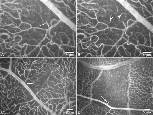
Fig. 2. Areas of capillary loss (no ADPase activity) in 31.2-year-old diabetic monkey 13 (A–C) and a 24.3-year-old diabetic monkey (D). Capillaries lacking ADPase activity (arrow) were present in the superficial retinal vasculature (A, C) and in the deep capillary network (arrowheads), when visualized with focus in two planes. Arteriolar pruning (arrow) was apparent at the edge of this area of capillary dropout. Lead ADPase reaction product with dark-field illumination.
Tel : 028-8592-1823 (in China)
Tel:+1-517-388-6508 (in US)
Tel : +86 (28) 8592-1823 (outside of China)
Fax : +86-28-62491302
Zip code : 610041
Address: No.88, Keyuan South Road, Hi-tech Zone, Chengdu, Sichuan Province, China